
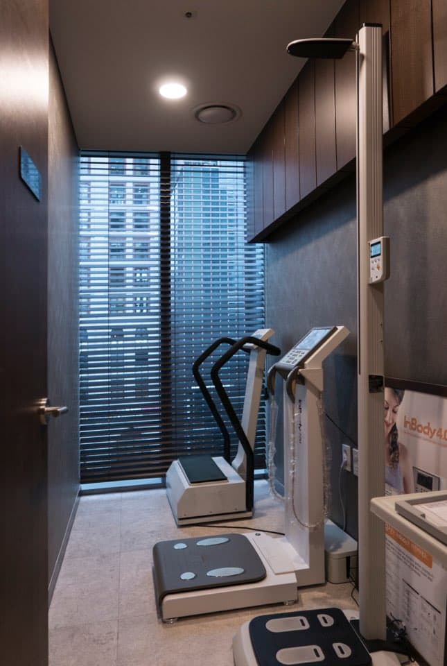
Ultrasound Analysis
Ultrasound-Based Analysis
Before surgery, ultrasound is used to check the thickness and boundary of the fat layer and the position, boundary and depth of muscle. Blood vessels are checked with Doppler when needed, and the findings inform the plan.
The Core of Ultrasound Analysis
Fat-Layer Depth Measurement
Ultrasound is used to check the thickness and boundary of the fat layer in each area.
Muscle position, boundary and depth
Ultrasound is used to check the position, boundary and depth of the muscles around the surgical area.
Vessel check, when needed
Where clinically needed, Doppler is used to check the position of major blood vessels, and this informs the surgical plan.
Customized Design
Based on the analysis, we build an individualized treatment plan for each person.
What ultrasound shows in consultation — and what it cannot settle
The ultrasound scan is carried out in the ultrasound examination room during the course of care. We check the thickness of the skin and subcutaneous fat, the boundary between layers and how fat is distributed, and the imaging findings are read together with the clinical examination.
When needed, ultrasound helps check skin and subcutaneous fat thickness, the boundary between layers, and fat distribution. It is not a device that definitively diagnoses every adhesion or predicts the result in advance. Measurements can vary with examination conditions, so they are read together with the clinical examination.
- Skin and subcutaneous fat thickness — how thick the layer is at the site being examined.
- The boundary between layers — where the fat layer separates from the structures beneath it.
- Fat distribution — how thickness varies within one area.
- Muscle position, boundary and depth — ultrasound is used to check the position, boundary and depth of the muscles around the surgical area.
What ultrasound does not decide
Ultrasound does not settle the answer on its own. None of the four below is determined by ultrasound alone; each is judged together with the clinical examination.
- It does not fix the outcome in advance — a measurement describes the present state, not how the result will look after surgery.
- Measurements can vary with examination conditions — probe pressure, the exact site measured and the examiner all change the value.
- Clinically agreed cut-off values do not yet exist — there is no established general rule that a thickness above some number of millimetres means surgery is indicated.
- It does not accurately replace whole-body fat percentage — whole-body estimates derived from site thicknesses are less accurate, particularly in overweight and obese people.
This page provides general medical information. It is not an individual diagnosis or a recommendation of treatment. Whether and how any method applies is decided after your current condition is examined in consultation.
References
The studies below support the explanations on this page. All are research from other institutions and are not treatment outcomes of NB Plastic Surgery Clinic.
- Neagu M, Neagu A. A Decade of Progress in Ultrasound Assessments of Subcutaneous and Total Body Fat: A Scoping Review. Life (Basel), 2025;15(2):236.
Supports · A paper describing the general evidence for, and the limits of, measuring subcutaneous fat thickness with ultrasound. What NB actually assesses follows the clinical scope set out on this page.
- Azzi AJ, Lafrenière AS, Gilardino M, Hemmerling T. Ultrasonography Technique in Abdominal Subcutaneous Adipose Tissue Measurement: A Systematic Review. Journal of Ultrasound in Medicine, 2019.
Supports · Supports the limit that measurements vary with probe pressure, measurement site and examiner, and that a standardised measurement protocol is needed.
Not sure yet? Start with a consultation.
See real procedure results.
Wondering which procedure suits you?
Our lead surgeon analyzes with ultrasound and designs a personalized plan.
Sinsa Stn, Gangnam, Seoul · Private parking tower
Mon–Fri 10:00–19:00 · Sat 10:00–16:00